Here is the cleaned up and expanded WordPress blog post:
Mushroom Photography Techniques: Capturing Microscopic Beauty
Introduction: The Hidden World Revealed Through the Lens
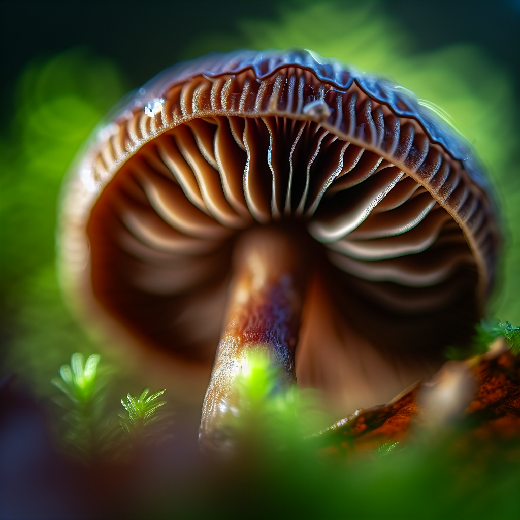
The mysterious world of mushrooms has captivated scientists, artists, and wellness enthusiasts for centuries. Apart from their culinary and medicinal uses, mushrooms possess an extraordinary aesthetic appeal—especially under microscopic observation. Mushroom photography, particularly when harnessed for medical and scientific purposes, offers a deeper look into the intricate structures that form the foundation of these fascinating fungi. From the delicate filigree of gill networks to the sculptural intensity of spores seen through magnification, the art and science of capturing mushrooms under a lens reveal an enchanting realm unseen by the naked eye.
In recent years, mushrooms have taken center stage in innovative natural health treatments, prominently featuring in areas like nootropic enhancement, anxiety relief, and even immunological support. These health benefits have spurred increased interest in studying the structure and composition of different mushroom species. As such, photography is not only a method of artistic expression but also a critical tool in advancing medical mycology and natural medicine research.
Mushroom photography blends technical skill with scientific curiosity. Whether for documenting rare species, observing disease-fighting compounds like beta-glucans, or creating awareness about sustainable remedies, specialized photography enhances our insight into the medicinal potential of mushrooms. With the rise of platforms like ShroomFan.com, enthusiasts and professionals alike are turning to high-resolution photography to share discoveries, diagnose fungal diseases, and promote therapeutic uses of psilocybin and other functional mushrooms.
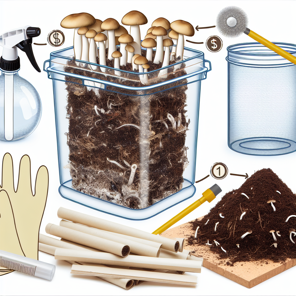
Capturing mushrooms—especially via macro and micro techniques—requires a unique photographic approach. Lighting, focal stacking, lens adaptation, and, in many cases, even laboratory equipment become necessary tools. Understanding fungal biology can also enhance the effectiveness of your visual storytelling, as different species demand varying conditions for optimal display.
This article will explore essential mushroom photography techniques, discuss their role in advancing medical studies and natural treatments, and provide insightful methods to harness technology in showcasing the breathtaking, microscopic world of mushrooms.
Illuminating the Invisible: How Photography Fuels Scientific Discovery
Mushroom photography has evolved beyond hobbyist interest into a pivotal element of scientific study, particularly in the field of fungal biology and integrative medicine. With the global rise in natural therapy applications, mushrooms are being examined more closely for their structural features, which often require crisp, detailed images for accurate classification and compound analysis.
One primary area where mushroom photography contributes to scientific progress is in taxonomy. According to a peer-reviewed study published in Mycologia, detailed photographic documentation, including microstructures like spores and basidia, significantly aids in identifying and verifying new fungal species. This is vital as certain medicinal fungi—like Ganoderma lucidum (Reishi), Hericium erinaceus (Lion’s Mane), and Psilocybe cubensis—share morphological similarities with toxic or less effective species. High-resolution imaging helps prevent misidentification and ensures safe usage in medical treatments.
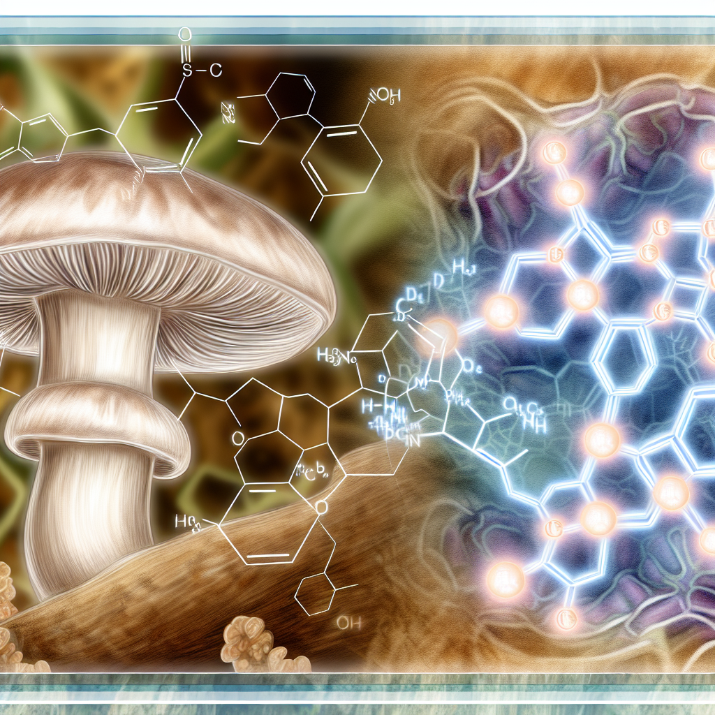
From Spore to Supplement: Visualizing Bioactive Compounds
Another crucial application is in the study of active compounds like polysaccharides, triterpenoids, and psilocybin. Research published by the National Institutes of Health (NIH) demonstrates how microscopic photography aids in tracking the morphology of mushrooms during various stages of growth, which aligns with the fluctuation of compound concentrations. For instance, understanding how cap formation correlates with peak psilocybin levels can inform harvesting schedules for researchers and medicinal cultivators.
Moreover, mycologists often use scanning electron microscopy (SEM) to visualize cellular-level components in mushrooms. This facilitates the understanding of spore distribution, defense mechanisms, and enzymatic factors involved in the fungi’s interaction with human biochemistry. These insights trickle down to real-world applications, such as improving mushroom supplement potency and validating claims made by companies marketing natural cures.
Tools of the Trade: Photographic Techniques and Technology
Practically speaking, innovations in imaging technology—from focus stacking in DSLR setups to the use of portable USB microscopes—have made high-quality imaging accessible for both labs and independent practitioners. The advancement of photogrammetry, which builds 3D images from multiple angle shots, also opens new avenues for virtual mushroom identification and medical research collaboration.
Key techniques in mushroom microscopy include:
– Focus Stacking – Combining multiple shots at different focus depths to create ultra-sharp images.
– Darkfield/Backlighting – Enhances the silhouette of microstructures.
– SEM (Scanning Electron Microscopy) – For hyper-detailed visuals of spores, filaments, and cellular layers.
– Lens Adaptation – Retro lens mounting or use of microscope objectives on DSLR setups.
– Specimen Preparation – Dehydration, staining, or freezing to preserve structural integrity.
Whether you’re capturing the gilled underside of a Lion’s Mane or the radial mycelia of a Cordyceps species, the right gear and technique make all the difference.
Beyond Beauty: Building Biological Databases Through Photography
Mushroom photography contributes not merely by capturing beauty but by providing indispensable data that can be analyzed for therapeutic applications and pharmaceutical development. Platforms like FungiMap and Mushroom Observer provide vast collections of macro and micro images that are routinely consulted by researchers working in complementary medicine. This collaborative visual database model ensures global access to consistent, quality-assured content crucial to the development of safe and effective natural therapies.
Photographers and scientists alike contribute to a growing library of knowledge that supports everything from taxonomy and conservation to medical breakthroughs and consumer health products. In this light, photographing mushrooms becomes a responsibility as well as a creative pursuit.
Conclusion: Merging Art and Science for a Healthier Future
Microscopic mushroom photography is more than just a visually stunning endeavor—it’s a critical bridge connecting mycology, medicine, and holistic wellness. As technology continues to evolve alongside our understanding of fungi, the use of precise, well-executed photography will remain an indispensable part of both artistic exploration and scientific innovation.
Whether you’re a passionate herbalist or a professional researcher, embracing mushroom microscopy allows for greater appreciation and more effective utilization of the ancient healing wisdom hidden within the fungi kingdom.
References
– Mycologia Journal, American Mycological Society
– National Institutes of Health – Medicinal Mushrooms in Preventive and Therapeutic Medicine
– Mushroom Observer – Community Mushroom Photography Database
– FungiMap – Enhancing Fungus Conservation through Photography
– NIH Study on Psilocybin-containing Mushrooms
– Advances in Scanning Electron Microscopy for Fungal Biology
—
Summary: This article explores the essential techniques and technologies of mushroom photography, highlighting how it plays a crucial role in advancing scientific discovery, understanding medicinal compounds, and promoting natural health treatments. From macro and micro imaging to cutting-edge 3D modeling, the article showcases the versatility of this field in bridging the gap between art, science, and holistic wellness.

Dominic E. is a passionate filmmaker navigating the exciting intersection of art and science. By day, he delves into the complexities of the human body as a full-time medical writer, meticulously translating intricate medical concepts into accessible and engaging narratives. By night, he explores the boundless realm of cinematic storytelling, crafting narratives that evoke emotion and challenge perspectives. Film Student and Full-time Medical Writer for ContentVendor.com